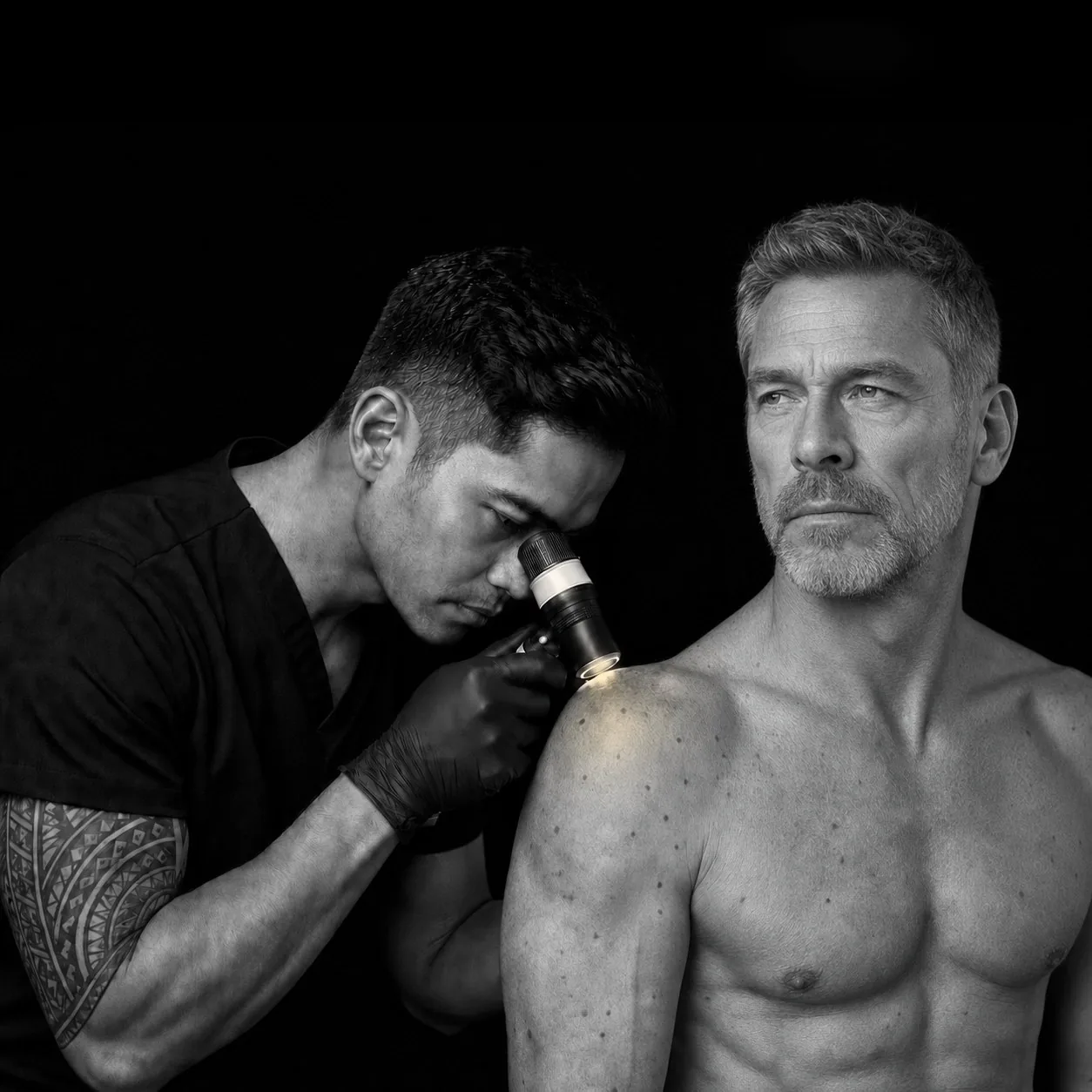
The One Sentence That Matters
I took a full day course in June 2026 called Basic Dermoscopy: Lesion Safety and Dermoscopy for Cosmetic Practitioners, run by SpotCheck and taught by Dr Ayub Khan, a GP and dermatology specialist who does mole mapping in the UK.
One slide said the whole thing:
"We are not here to teach you how to make a diagnosis. We are here to teach you when to stop, check and refer."
That is the honest description of what I do with a dermatoscope, and it is the frame for everything below.
What a Dermatoscope Actually Is
A dermatoscope is a lens with a light in it, held against the skin, usually at ten to fourteen times magnification.
The interesting part is the light rather than the magnification.
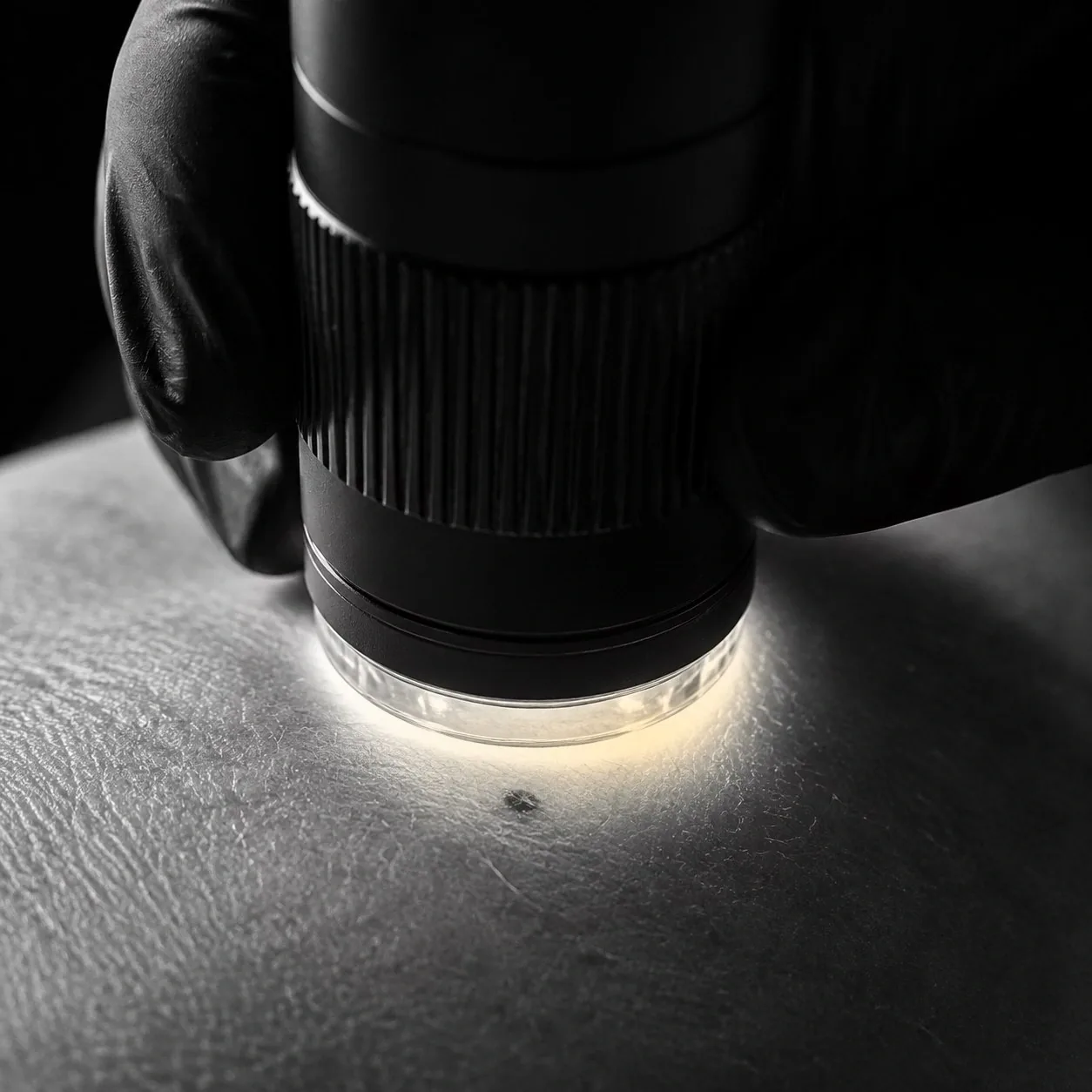
Skin has a problem, which is that the surface layer reflects and scatters most of the light hitting it. That glare is what stops you seeing anything underneath. Magnify glare and all you get is bigger glare.
Two ways around it:
- contact, with fluid. A glass plate and a drop of oil, alcohol or gel fill the microscopic air gaps in the surface so light goes in instead of bouncing off. This shows the top layers down to the junction between epidermis and dermis
- polarised. Two filters crossed at ninety degrees block the light that bounced straight off the surface and let through only light that scattered inside the skin. This reaches deeper, into the upper dermis, and needs no contact and no fluid
They show different things, which is why good devices do both. Polarised light shows blood vessels, reds and pinks, and white shiny structures. Non-polarised shows blue-white colours, milia-like cysts and comedo-like openings, and works better on palms and soles.
This is also the difference between a dermatoscope and a clip-on macro lens for a phone. The phone lens gives magnification and nothing else. A dermatoscope gives controlled light at the right angle, cross-polarisation to defeat the glare, a fixed working distance so focus and scale repeat from visit to visit, and usually a reticle so a lesion can actually be measured rather than estimated.
What It Shows That the Eye Cannot
Under the light, skin stops being a colour and becomes a structure. The features that matter have names:
- pigment network, a honeycomb grid of brown lines. Even and regular is ordinary. Irregular holes and thickened lines are not
- dots and globules, which tell you the lesion is melanocytic. Uneven size, shape and scatter is the version that gets attention
- streaks and pseudopods at the edge, meaning growth that is pushing outward in one direction rather than all directions
- a blue-white veil, a structureless area of confluent blue
- regression, scar-like depigmentation where the immune system has attacked the lesion. This shows up in roughly half of melanomas
- vessel patterns. Dotted, linear and corkscrew vessels each mean something different
- five or six colours in one lesion rather than one or two
None of that is visible at arm's length. Most of it is not visible at close range either.
Clinical Symmetry Is Not Dermoscopic Symmetry
This was the slide that reorganised how I look at skin.
A lesion that looks perfectly even to the naked eye can be chaotic under the scope. A lesion with a ragged outline can be completely orderly underneath.
The two do not track each other, in either direction. The course made the point with four moles that looked essentially identical unmagnified and turned out to be four different things once the light went on.
That is the argument against assessing anything by eye alone. The naked-eye rules everyone knows are a starting point, not a filter.
Why This Matters Before Anyone Treats a Skin Lesion
Here is where it stops being academic.
A large part of the cosmetic industry removes small skin lesions with electrosurgery. Skin tags, seborrhoeic keratoses, the raised bumps people want gone. It is fast, it is cheap, and it is popular.
The problem is the look-alikes. From the course, unmagnified and side by side:
- sebaceous hyperplasia and nodular basal cell carcinoma. Under the scope one has white clods and crown vessels, the other has arborising vessels and blue-grey globules
- basal cell and squamous cell carcinoma. Fine arborising vessels against white circles and dotted vessels
- cherry angioma and pyogenic granuloma, then pyogenic granuloma and squamous cell carcinoma
- seborrhoeic keratosis and melanoma. Comedo-like openings and milia-like cysts against streaks, grey-white areas and chaos
Every one of those pairs is a benign thing a clinic would happily treat sitting next to something that needs a doctor.
The course showed a case treated by an aesthetic clinic with electrosurgery, before and after. The lesion came back, and it came back looking considerably worse than it went in. Burning off a lesion nobody identified does not just fail. It destroys the evidence, and the thing that grows back is harder to assess than the thing that was there.
That is the risk in one picture, and it is why the training exists.
The Evidence, Including the Part That Cuts Against Me
The numbers for dermoscopy are good.
The Cochrane review covering 104 studies and nearly 43,000 lesions found that in-person dermoscopy reached 92 percent sensitivity for melanoma against 76 percent for looking with the naked eye, at matched specificity. Per thousand lesions examined, that is nineteen additional melanomas found without any increase in false alarms.
Now the part that matters more.
A 2002 review in Lancet Oncology of 27 studies found dermoscopy clearly beat the naked eye overall, and then found this: dermoscopy performed by untrained or less experienced examiners was no better than clinical inspection without dermoscopy at all.
The device does nothing on its own. Handed to someone untrained it produces confidence rather than accuracy, which is worse than no device, because confidence is what stops people referring.
I would rather publish that sentence than leave it out. It is the reason I did the course before touching the subject, and it is the question worth asking anyone in this industry who mentions dermoscopy: what training, and when. I have written before about what my qualifications actually cover.
Why an Esthetician Often Sees It First
The argument here is about access, not expertise, and I want to be careful about the difference.
I look at skin that people cannot see themselves, under good light, for forty-five minutes at a time, on the same body repeatedly over months or years. A family doctor gets ten minutes and a shirt that stays on.
The research on this is real but narrow:
- a 2024 study trained 355 estheticians. Before training, 68 percent had not recommended a single client see a doctor about a suspicious lesion in the previous three months. After training, 87 percent felt very comfortable doing so. The authors called estheticians a valuable screening partner for dermatologists
- a 2025 UK study of hair professionals found only 5 percent had any formal skin cancer training, yet 35 percent had advised a customer about a suspicious lesion. Of those, 39 percent said the customer went on to receive a skin cancer diagnosis
- there are published cases of scalp melanomas found by hairdressers, on a part of the body nobody can examine themselves
What none of those studies show is that we are accurate. They measure comfort, willingness and self-reported outcomes. The honest version of the claim is that I am in the room, often, with the light on, looking at skin you cannot see. That is worth something. It is not a screening programme.
What I Am Allowed to Say, and What I Am Not
Here is a detail worth knowing about my own training: the course was taught under UK law, by a UK doctor, to a room that was mostly Canadian. His scope of practice slide said so in those words. UK rules are not our rules, so rather than assume the training covered me, I went and read the Ontario statute myself.
Estheticians and electrologists are not regulated health professionals in Ontario. We are overseen as personal service settings under the Health Protection and Promotion Act and inspected by the local board of health, which is public health oversight rather than clinical scope. I have written about what the regulations actually cover here.
The relevant law is the Regulated Health Professions Act, 1991. Section 27(2) lists the controlled acts, and the first one reads:
"Communicating to the individual or his or her personal representative a diagnosis identifying a disease or disorder as the cause of symptoms of the individual in circumstances in which it is reasonably foreseeable that the individual or his or her personal representative will rely on the diagnosis."
Read that carefully, because the restricted act is the communicating, not the looking.
Nothing on that list covers magnifying or illuminating intact skin. The second controlled act is performing a procedure below the dermis, and a dermatoscope does not break the surface. The seventh is applying a form of energy prescribed by regulation, and the prescribed list is specific and short: electricity for ten named medical purposes, electromagnetism for MRI, soundwaves for ultrasound and lithotripsy. Light from a lens is not on it.
There is also section 30, which prohibits anyone outside their scope from treating or advising on health where serious bodily harm is reasonably foreseeable.
So the line runs through my mouth rather than my hands. In practice:
- what I can say: "there is a spot here that does not look like your others, and I cannot tell you what it is. Please have your doctor or a dermatologist look at it."
- what I cannot say: "that looks like a melanoma."
- what I equally cannot say: "that is nothing, do not worry about it."
That third one catches people out, including practitioners. Reassurance is a diagnosis. It is the more dangerous of the two, because the person who has been alarmed goes to a doctor and the person who has been reassured does not.
So the limit runs in both directions. I cannot tell you what something is, and I cannot tell you it is nothing. What I can do is tell you that it does not look like your others, and that somebody qualified should settle it.
This Is Not a Service I Sell
I want to be plain about this, because a post like this can read as a soft advertisement and it is not one.
There is no skin check on my price list. No mole mapping, no lesion screening, no dermoscopy appointment to book. What there is, is somebody who looks carefully at skin during hair removal and who knows a great deal more than he did in May about when to stop and say something.
Whether this can properly be sold as a service in Ontario is a question I do not think is settled, and until it is, I am not charging for it. If that changes, I will say so here.
What It Cannot Do
A dermatoscope has real blind spots, and you should know them:
- it is not a diagnosis. Everything above is measured against biopsy, which is the only thing that settles the question
- nodular melanoma grows down rather than out, and a third of them carry no pigment at all. They can fail every visual rule and look unremarkable under the scope
- amelanotic melanoma, between 2 and 20 percent of all melanomas, is difficult by dermoscopy and often found late for exactly that reason
- it sees the upper dermis and no further
- it cannot see skin I am not looking at, and it knows nothing about how long something has been changing. You know that part. Tell me
It also does not tell you what an ordinary irritated follicle is. If what you are worried about arrived after hair removal rather than before it, the more likely answers are covered in the pieces on telling bumps apart and folliculitis.
The Number That Explains the Urgency
Melanoma in Canada is estimated at 11,300 new diagnoses and 1,250 deaths this year. Overall five-year survival is 90 percent, which sounds reassuring until it is broken out by stage.
Five-year melanoma-specific survival runs 99 percent at stage 1A and 32 percent at stage 3D.
That gap is the entire argument. Nothing I do moves that number. Saying something early, to the right person, does.
Bottom Line
Dermoscopy is a light and a lens that let someone see structure instead of colour, and it changes what is visible on skin in a way that magnification alone does not.
It does not make me a diagnostician. Ontario law is clear that naming a disease is not mine to do, and the evidence is equally clear that an untrained person holding a dermatoscope performs no better than one without it.
What it makes me is a better referrer. I see skin you cannot see, regularly, under good light, and I know enough now to recognise when something does not belong and to say so without pretending to know what it is. That cuts both ways. I will not tell you something is cancer, and I will not tell you it is nothing.
If I ever tell you to get something looked at, I am not diagnosing you and I am not trying to frighten you. I am doing the one useful thing available to me, which is stopping, checking, and sending you to someone who can settle it.
Where I use it most deliberately. Surgical donor site work means assessing a mapped rectangle of skin on a trans man before eighteen months of treatment go into it, moles, scars and keloid history included. That assessment is described in the donor site guide.
If this is your situation, these go together. hidradenitis suppurativa and hair removal, the pain map, every area ranked, how often you should actually exfoliate, tattoos and hair removal and burns, hyperpigmentation and scarring, and how to avoid them.